PennHip Xray For Dogs In Mt Pleasant SC
An advanced, multi-faceted radiographic screening method for canine hip evaluation.
![]()
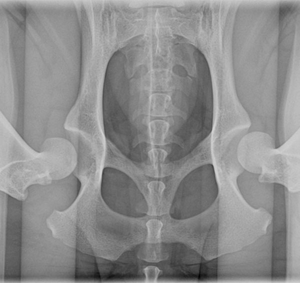
What is PennHip? PennHip is an x-ray of your dog’s hip joints which is commonly used to determine if your dog has hip dysplasia or is at risk of hip dysplasia.
Need a PennHip xray for dogs in Mt Pleasant SC? Mount Pleasant Animal Hospital offers PennHip xrays for dogs: a multifaceted radiographic screening method for canine hip evaluation. This technique assesses the quality of the dog’s hips and quantitatively measures canine hip joint laxity. It is used to screen dogs for canine hip dysplasia and osteoarthritis, diseases that cause pain and disability in dogs who have it.
Upon completion of PennHip radiographs by a certified veterinarian, the images are sent to the PennHip Analysis Center for evaluation. Pet owners then receive a full report explaining the degree of hip laxity found in their dog, an analysis of how their dog’s hips compare to the average dog of their breed, and what they can expect to see moving forward regarding hip pain and disability. If hip dysplasia is detected, information regarding how to best protect their dog’s hips moving forward also is provided. If you’re concerned about your dog’s hips, Mount Pleasant Animal Hospital provides services for a PennHip xray for dogs in Mt Pleasant SC.
What advantage does a PennHip xray for dogs have compared to OFA Radiographs? (Orthopedic Foundation of America)
Traditional hip screening imaging (OFA radiographs) consists of a single radiographic view: the hip extended view, otherwise known as the “OFA view.” This single view is used to obtain information regarding the existence of osteoarthritis (OA) of the hip joint. This view is subjectively scored for osteoarthritis (which develops later in the disease process) and often cannot assess the underlying joint laxity that is causing the arthritis. Due to the often late development of visible disease, OFA radiographs are recommended to be taken only in dogs 2 years of age or older.
In contrast, PennHip radiographs consist of 3 views: the distraction view, the compression view, and the hip-extended view. The distraction view and compression view are used to obtain accurate and precise measurements of joint laxity and congruity, respectively. The hip-extension view also is reviewed to obtain supplementary information regarding any existing osteoarthritis. Due to these additional, quantitative measurements, canine hip dysplasia can be detected much earlier. PennHip xrays for dogs can be performed in dogs as early as 16 weeks old.
What advantage does it have over OFA (Orthopedic Foundation of America)
Traditional hip screening imaging (OFA radiographs) consists of a single radiographic view: the hip extended view, otherwise known as the OFA view. This single view is used to obtain information regarding the existence of osteoarthritis (OA) of the hip joint. This view is subjectively scored for osteoarthritis (which develops later in the disease process) and often cannot assess the underlying joint laxity that is causing the arthritis. Due to the often late development of visible disease, OFA radiographs are recommended to be taken only in dogs 2 years of age or older.
PennHip radiographs consist of 3 views: the distraction view, the compression view and the hip-extended view. The distraction view and compression view are used to obtain accurate and precise measurements of joint laxity and congruity, respectively. The hip-extension view is also reviewed to obtain supplementary information regarding any existing OA. Due to these additional, quantitative measurements, canine hip dysplasia can be detected much earlier and can be performed in dogs as early as 16 weeks old.