In-House Laboratory

We are able to perform detailed testing that rapidly aids us in the diagnosis of your pet and determination of the best Our specifically trained Laboratory Technician performs these diagnostic tests for our veterinarians while your pet is being treated, whether on a hospitalized basis or as an outpatient. treatments.
[expand title="Read More" swaptitle="Read Less" trigclass="arrowright" targpos="inline" targtag="span" trigpos="below"]
A few of the many important tests we provide include:
For those few tests that we do not perform at our clinic, we use an outside laboratory, Antech Diagnostics Labs. They are the nation’s premier veterinary diagnostics lab and provide us rapid testing and reporting on possible diseases, providing:
Remember pets age approximately 7 years for every single calendar year. This is why annual or bi-annual blood screening is important. Some conditions can thus be detected before serious illness becomes clinically apparent. We also provide tailored testing to your pet’s age, lifestyle, and breed. During your pet’s next wellness exam ask us about the screening tests that are right for you and your pet.
[/expand]
- Blood analysis, complete blood counts, blood chemistries
- Urinalysis and sedimentation
- Fecal examinations (testing for the presence of hookworms, whipworms, tapeworms, and roundworms)
- Viral identification (Parvovirus, Feline Leukemia Virus, and Feline Immunodeficiency Virus)
- Heartworm disease testing
- Skin scrapes
- Skin cytology, ear cytology, mass cytology
- Bacterial cultures
- PCR panels
- Comprehensive thyroid panels
- Fluid cytology
- Fungal cultures
- Histopathology
- Tick panels
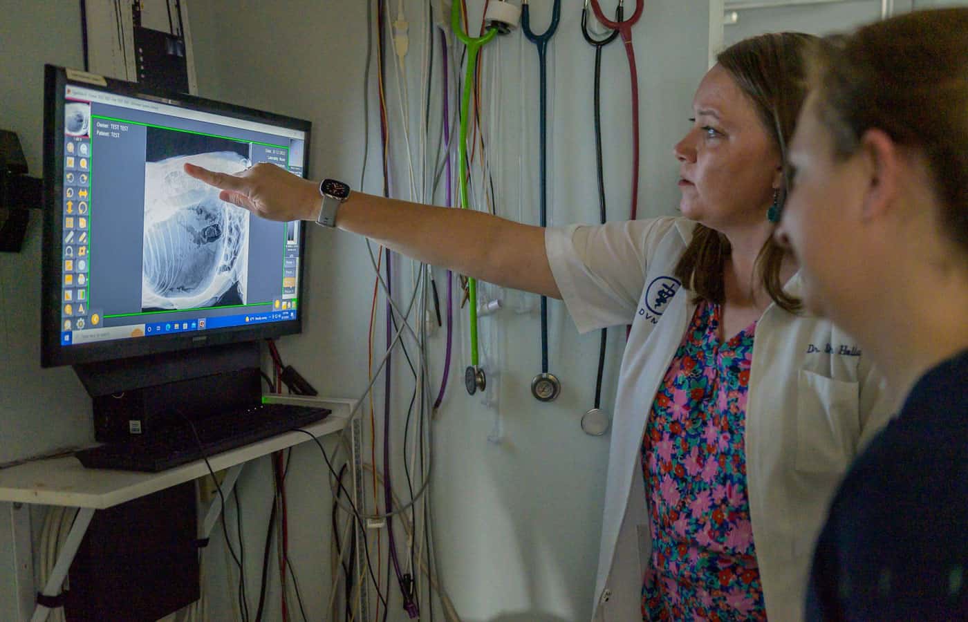
X-Rays

Digital Radiology/Radiographs (X-Rays) are used to diagnose various internal problems such as:
[expand title="Read More" swaptitle="Read Less" trigclass="arrowright" targpos="inline" targtag="span" trigpos="below"]
 OFA Certification also requires X-Ray images of the hips and elbows to assess joint laxity.
The images are obtained digitally for immediate interpretation by our dedicated veterinarian. Digital images are also reviewed via telemedicine by board-certified radiologists (Oncura). The patient rarely needs sedation but may depending on the number of pictures required, restraint needed for proper positioning, or if the images are needed for OFA Certification. In general, X-Rays are painless and non-invasive and an excellent way to help diagnosis illness and injury.
Mount Pleasant Animal Hospital has digital pet dental and full-body radiography capabilities, utilizing the latest technology to produce instant images of the skull region in extreme detail.
Penn-Hip X-rays
[/expand]
- Bone Fractures
- Bladder Stones
- Mass/Tumor Detection
- Swallowed Items or Lodged Foreign Objects
 OFA Certification also requires X-Ray images of the hips and elbows to assess joint laxity.
The images are obtained digitally for immediate interpretation by our dedicated veterinarian. Digital images are also reviewed via telemedicine by board-certified radiologists (Oncura). The patient rarely needs sedation but may depending on the number of pictures required, restraint needed for proper positioning, or if the images are needed for OFA Certification. In general, X-Rays are painless and non-invasive and an excellent way to help diagnosis illness and injury.
Mount Pleasant Animal Hospital has digital pet dental and full-body radiography capabilities, utilizing the latest technology to produce instant images of the skull region in extreme detail.
Penn-Hip X-rays
[/expand]
OFA Certification also requires X-Ray images of the hips and elbows to assess joint laxity.
The images are obtained digitally for immediate interpretation by our dedicated veterinarian. Digital images are also reviewed via telemedicine by board-certified radiologists (Oncura). The patient rarely needs sedation but may depending on the number of pictures required, restraint needed for proper positioning, or if the images are needed for OFA Certification. In general, X-Rays are painless and non-invasive and an excellent way to help diagnosis illness and injury.
Mount Pleasant Animal Hospital has digital pet dental and full-body radiography capabilities, utilizing the latest technology to produce instant images of the skull region in extreme detail.
Penn-Hip X-rays
[/expand]Ultrasound

Ultrasound is a non-painful, non-invasive diagnostic technique used in both animal and human medicine. As in human ultrasound imaging,this technique uses a lubricating gel and probe that passes over the abdomen and chest regions of the patient to assess soft tissues and detect diseases, cancers,
[expand title="Read More" swaptitle="Read Less" trigclass="arrowright" targpos="inline" targtag="span" trigpos="below"] and pregnancy. Unlike X-Rays, ultrasound uses sound waves to penetrate tissues, producing real-time images/videos of the body cavity or organ systems and their functioning.
When the heart is the target of study, it is called an echocardiogram or echo. The series of images obtained from an echo can be used to interpret a physical exam finding of a heart murmur or rhythm disturbance, stage cardiac disease, or uncover a congenital defect of the heart.
Ultrasound can also be used to explore other areas of the body to aid in:
Rarely does an ultrasound procedure itself require sedation as it is non-invasive and painless. When an ultrasound is performed, a small target area of hair may be shaved in order to enhance the contact between the probe and the skin. These decisions are discussed with you ahead of time to insure the best plan for you and your pet.
For your convenience and ours, we offer two options to our patients undergoing ultrasound procedures. You may choose to drop off your pet or wait while we take, process, and interpret the images.
Dr. Kevin Shuler has advanced training in echocardiology and abdominal ultrasonography. In addition, we do consult with Board Certified cardiologists and radiologists through telemedicine (Oncura) and the local referral centers of Veterinary Medical Care or Charleston Veterinary Referral Center.
[/expand]
- A Needle-guided Sample Collection
- Detecting Organ Disease (such as liver, kidney, or bladder)
- Tumor/Mass Detection


